

Penile Anatomy and Tunical Architecture
The human penis consists of intricate concentric structural layers, extending from the superficial skin and dartos fascia, Buck’s fascia, and neurovascular bundles, down to the thick bilaminar tunica albuginea and the cavernous erectile tissue. These tissues possess physiological elasticity and compliance required for erection and elongation.
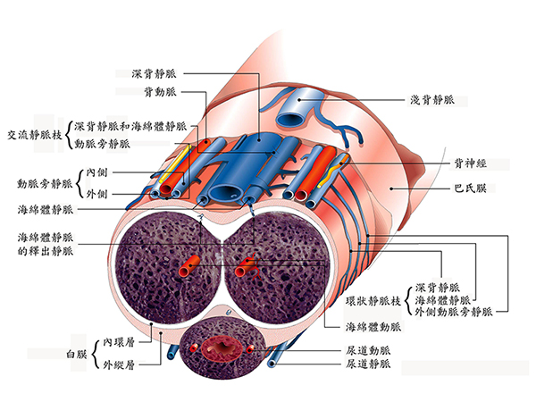
Penile curvature becomes clinically prominent upon erection and is categorized into congenital and acquired forms. While congenital curvature stems from disproportionate tunical growth without palpable scarring, acquired penile curvature is predominantly caused by Peyronie’s disease—a condition characterized by aberrant wound healing and excessive collagenous plaque formation in the tunica albuginea following microvascular or mechanical trauma.
Etiology and Pathophysiology
Peyronie’s disease affects approximately 5% to 9% of men over 40 years of age. Repetitive microvascular bending or buckling trauma during sexual intercourse causes micro-tears at the junction between the tunica albuginea and cavernous septa. Instead of normal tissue remodeling, an excessive fibrous plaque develops. Because the plaque lacks physiological elasticity, it restricts expansion during erection, producing a hinge effect, severe lateral/dorsal angulation, penile shortening, or hourglass waist deformities (Figure 2).


Figure 2: Tunical plaque formation in Peyronie’s disease. A) Dorsal plaque producing an acute upward hinge bend. B) Circumferential waist narrowing resulting in an hourglass deformity.
Patients often report a history of an acute buckling sensation or sudden pain during vigorous sexual activity. Associated medical conditions include Dupuytren’s contracture (palmar fascial fibromatosis), plantar fasciitis, diabetes mellitus, and systemic vascular disease. Approximately 37% to 58% of men with Peyronie’s disease suffer from coexisting erectile dysfunction, frequently attributable to secondary penile venous leakage.
Clinical Signs and Diagnostic Evaluation
Physical palpation along the penile shaft typically reveals a well-demarcated fibrous nodule or thickened band, most frequently on the dorsal aspect. In advanced cases with dystrophic calcification, the plaque can feel stony hard. Comprehensive evaluation includes:
- Detailed medical and sexual history, documenting onset and progression.
- Multi-angle photographic documentation taken during full erection at home.
- Penile Color Doppler Duplex Ultrasound to assess plaque dimensions, calcification, and vascular hemodynamics (peak systolic velocity and end-diastolic velocity).
Treatment Phases and Surgical Timing
Peyronie’s disease typically follows a biphasic clinical course: an active (acute inflammatory) phase lasting 6 to 12 months, followed by a stable (chronic) phase. Conservative non-surgical management is strictly indicated when:
- Active penile pain persists during erection or flaccidity.
- The plaque or curvature is actively evolving and progressing.
- The disease duration is under 12 months without documented anatomical stabilization.
Surgical reconstructive correction (such as tunical shortening or tunical grafting) should only be considered when the deformity has remained stable and pain-free for at least 6 months, and when curvature precludes satisfactory sexual intercourse. Thorough clinical assessment by an experienced urologist is essential.
Clinical article authored and reviewed by Dr. Cheng-Hsing Hsieh.